
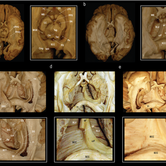
Ambiguity surrounds the existence and morphology of the human forniceal commissure. We combine advanced in-vivo tractography, multidirectional ex-vivo fiber dissection, and multiplanar histological analysis to characterize this structure’s anatomy.
Across all 178 subjects, in-vivo fiber dissection based on the Human Connectome Project 7 T MRI data identifies no interhemispheric connections between the crura fornicis. Multidirectional ex-vivo fiber dissection under the operating microscope demonstrates the psalterium as a thin soft-tissue membrane spanning between the right and left crus fornicis, but exposes no commissural fibers. Multiplanar histological analysis with myelin and Bielchowsky silver staining, however, visualizes delicate cruciform fibers extending between the crura fornicis, enclosed by connective tissue, the psalterium.
The human forniceal commissure is therefore much more delicate than previously described and presented in anatomical textbooks.
This finding is consistent with the observed phylogenetic trend of a reduction of the forniceal commissure in non-human primates compared to non-primate eutherian mammals.


Comments